Your dog’s expressive eyes show love, excitement, sadness, apprehension, and so many other emotions. If your beloved four-legged companion experiences eye problems however, he or she may not be able to communicate as well and could even suffer vision loss. It’s important to be able to recognise dog eye problems in their early stages because quick intervention can, in many cases, be the key to a better outcome.
STRUCTURE OF THE CANINE EYE
In learning to identify dog eye problems, it’s helpful to have an understanding of the unique structure of your dog’s eyes.
Like most mammals, your dog’s eyes are globe-shaped with two fluid-filled chambers, anterior and posterior. The chambers are separated by the lens, which is the structure that helps focus light beams onto the rear part of the eye, the retina. Your dog’s eye has a cornea, which is the outer, clear surface, which offers protection to the inner eye. Your dog’s eyes also have pupils and irises, like your eyes, muscles to help them move, open and close the lens, letting in an appropriate amount of light.
Your dog also has an upper and lower eyelid, like you, but one of the features that is unique to your dog is the existence of a prominent third eyelid, called the nictitating membrane. The third eyelid is located at the lower inner corner of the eye, between the lower eyelid and the eye. The third eyelid is thought to offer protection for the eyeball and to help in removing foreign bodies from the eye. There is also a gland located in the third eyelid which is believed to produce 30 percent of the aqueous part of your dog’s tear film. Normally, the third eyelid is concealed beneath the lower eyelids, but may become prominent in one or both of your dog’s eyes if there is a problem. For this reason, it’s a good idea to familiarise yourself with the anatomy of your own dog’s eyes including the position of the third eyelid.
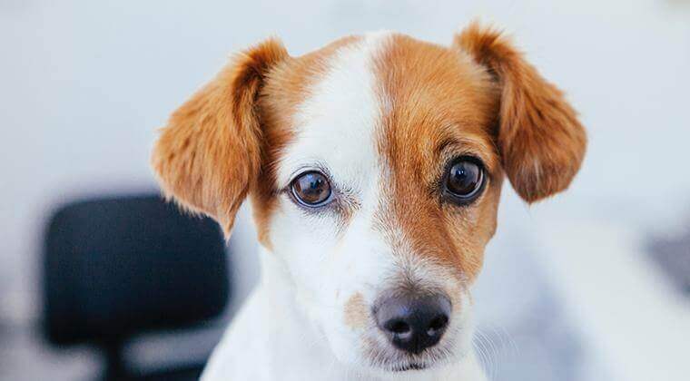
1. WHAT IS CHERRY EYE?
A condition that specifically affects the third eyelid, “cherry eye” gets its moniker from the red swollen mass that appears in the eye when a dog is stricken with this condition. Cherry eye in dogs occurs when the gland in the third eyelid thickens and slips out of its proper place.
You might occasionally get a glimpse of your dog’s third eyelid when he or she is sleeping or if your pet has had surgery and is recovering from anaesthesia. If your dog has cherry eye however, you might notice a red swelling protruding from the edge of the third eyelid, which is usually the first sign of this disease. The swelling may occur in one or both eyes. Some dogs may not have any other signs, while others may have signs akin to conjunctivitis.
Cherry eye is most commonly seen in dogs aged six months to two years.
It is not preventable because the cause is not known, but there may be a genetic predisposition. However, when you know what signs to look for, you can act quickly and get veterinary attention for your pooch.
Your vet plays a big role in your pet’s health. Enter your location and get a list of vets near you.
FIND A VETHOW IS CHERRY EYE IN DOGS TREATED?
Your vet may recommend a course of eye drops as the first step in treating cherry eye. If the cherry eye persists, the swelling does not decrease, and your dog is uncomfortable, surgery may be recommended.
2. CONJUNCTIVITIS
Conjunctivitis, or “pink eye,” is inflammation of the conjunctiva and is quite common in dogs. Signs include redness of the eye, swelling of the tissue around the cornea, discharge from the eye, and mild eye discomfort, with signs like keeping the eyes closed or rubbing them.
There are a wide range of causes of conjunctivitis in dogs such as allergies, injury, birth defects, and tear duct problems, to foreign matter, dry eye, distemper, or even tumours.
To treat conjunctivitis, your vet will first want to learn what’s causing it. Depending on the source of the inflammation, treatment can include the following procedures:
- Removal of the irritant and soothing the area with pain medication
- Prescribing antibiotics and saline washes to manage infection
- Medication for allergies
3. EPIPHORA
The condition of excessive tearing is known as “epiphora.” Signs of this dog eye problem include watery, teary eyes, which can result in stained or smelly hair below the eyes.
Epiphora in dogs can be the result of many conditions where there is excessive tear production or reduced tear drainage including abnormal eyelashes, conjunctivitis, and corneal ulcers.
Miniature and toy breeds seem more prone to epiphora. The most common cause of this is poor drainage of tears. Sometimes, a dog’s eyelids turn inward, which also irritates the eye and impedes the tear drainage system. Surgery may be an option to improve the drainage of tears or correct the eyelid position.
For the most part, your vet will want to find out what’s causing the epiphora and treat the problem appropriately.

4. DRY EYE
A dog with dry eye or keratoconjunctivitis sicca lacks the lubricating effect of tears, an issue that can lead to blindness if left untreated.
Dry eye often looks like recurrent conjunctivitis or persistent infection and can include a sticky, mucus discharge in the eyes, a dry nose and cloudiness of the cornea with or without visible ulcers.
The most common cause of dry eye is when the dog’s immune system attacks the tear glands.
Dry eye in dogs is diagnosed using a simple tear-measuring strip test to measure tear production that is painless and easy to do. Treatment for dry eye aims to restore tear production using medication that is applied to the eye twice daily. Some dogs may require supportive treatment with antibiotic drops or ointment and artificial tears.
5. GLAUCOMA
Glaucoma occurs when an imbalance in production and drainage of fluid in the eye (aqueous humor) causes a buildup of fluid that increases eye pressure to unhealthy levels. Not only can this be quite painful for your pooch, but it can be accompanied by redness of the eyes and vision loss.
Signs of glaucoma in dogs include dilated pupils (the pupil in one eye may be a different size from the other eye), redness of the eyes, or dilation of the blood vessels in the white part of the eye (the sclera).
There may also be a noticeable bulging of one or both eyes. Your dog may partially close his eyes or rub at the eyes, and you may notice lethargy in your pet, loss of appetite or even unresponsiveness. These signs can be due to the pain glaucoma can cause. If your dog displays any signs of glaucoma, he should be seen by a vet immediately to avoid vision loss.
Glaucoma in dogs is diagnosed using a tonometer, a special device to measure the pressure in the eye. Your vet will suggest appropriate medical and/or surgical treatment for your dog.

6. CATARACTS
A cataract occurs when the lens becomes cloudy or opaque, effectively blocking light from reaching the retina. This causes loss of vision ranging from mild vision problems to blindness. Cataracts should not be confused with the minor lens imperfections in young dogs or with the normal increase in the thickening and hardening of central lens tissue (sclerosis) that occurs in older animals.
The most common cause of cataracts in dogs is genetics (inherited).
Inherited cataracts can affect many breeds of dog, but some breeds seem to be more prone than others. Other causes include diabetes, malnutrition, radiation, inflammation, and trauma. In diabetes, the cataracts are due to glucose degradation products binding to lens proteins. Most diabetic dogs develop cataracts.
WILL MY DOG GO BLIND?
Cataracts do not mean certain blindness. In fact, dogs have such a keen sense of hearing and smell that vision loss may not be noticed. A cataract may also not block the vision entirely. Whether the cataract remains static or progresses depends on the type of cataract, the breed of your dog and other various risk factors.
Many dogs manage very well without specific therapy for their cataracts. Cataracts can be removed surgically by specialist veterinary ophthalmologists. You should speak to your vet to further evaluate your dog’s condition and evaluate appropriate treatment options.
No one likes to see their beloved pet suffer, so if you notice your dog pawing at his/her eyes, rubbing her face on the floor or carpet, you should be aware that this is a sign that there is a problem with her eyes. It’s a good idea to consult your vet at the first sign of an eye problem. In some cases, like glaucoma, this could save your dog’s vision.







 Go To United States
 Austria
 Belgium
 Czech Republic
 Denmark
 Europe
 Finland
 France
 Germany
 Greece
 Hungary
 Ireland
 Israel
 Italy
Italy Netherlands
 Norway
 Poland
 Portugal
 Romania
 Slovakia
 Spain
Spain Sweden
 Turkey
 United Kingdom